Figure 6
Trypanosoma dionisii as an experimental model to study anti-Trypanosoma cruzi drugs: A comparative analysis with benznidazole, posaconazole and amiodarone
De Souza W*, Barrias ES and Borges TR
Published: 17 October, 2018 | Volume 1 - Issue 1 | Pages: 014-023
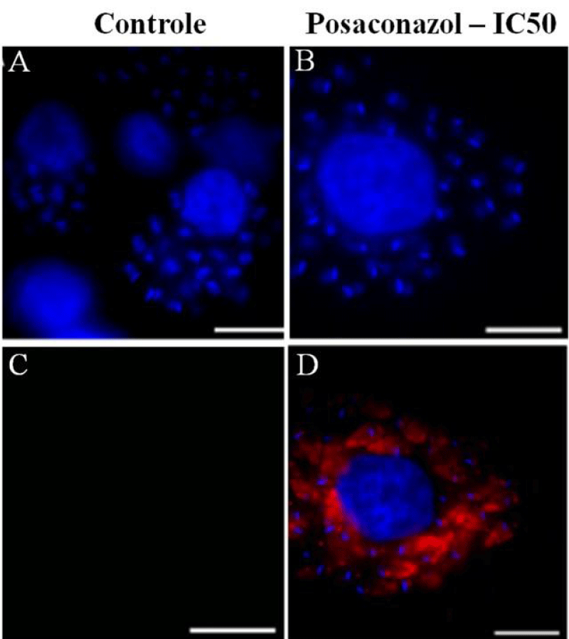
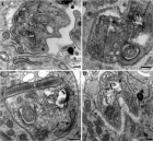
Figure 6:
Immunofluorescence microscopy demonstrating labeling of structures when cells were incubated in the presence of antibodies against LC3B (microtubule-associated protein light chain 3), a marker of autophagic cell death. Labeling was seen in intracellular amastigotes treated with posaconazole. (A–C) Untreated infected macrophages after 96 h of incubation are not labeled. (D–F) Intracellular amastigotes treated with 1 nM posaconazole exhibit intense red labeling (arrowhead). Bar = 5 µm.
Read Full Article HTML DOI: 10.29328/journal.ijcmbt.1001003 Cite this Article Read Full Article PDF
More Images
Similar Articles
-
Trypanosoma dionisii as an experimental model to study anti-Trypanosoma cruzi drugs: A comparative analysis with benznidazole, posaconazole and amiodaroneDe Souza W*,Barrias ES,Borges TR. Trypanosoma dionisii as an experimental model to study anti-Trypanosoma cruzi drugs: A comparative analysis with benznidazole, posaconazole and amiodarone. . 2018 doi: 10.29328/journal.ijcmbt.1001003; 1: 014-023
Recently Viewed
-
The effect of frequency of sexual intercourse on coronary artery diseaseMehdi Karasu*,Özkan Karaca,Mehmet Ali Kobat,Tarık Kıvrak,Mehmet İkbal İpek. The effect of frequency of sexual intercourse on coronary artery disease. Arch Vas Med. 2022: doi: 10.29328/journal.avm.1001015; 6: 001-004
-
A Case Report on Paradoxical EmboliYou Li* and Jason Wheeler. A Case Report on Paradoxical Emboli. Arch Vas Med. 2024: doi: 10.29328/journal.avm.1001019; 8: 004-007
-
Precision and personalized vaccines needed to face COVID-19 pandemicDahmani Fathallah M*. Precision and personalized vaccines needed to face COVID-19 pandemic. Insights Clin Cell Immunol. 2021: doi: 10.29328/journal.icci.1001017; 5: 003-003
-
NAD⁺ Biology in Ageing and Chronic Disease: Mechanisms and Evidence across Skin, Fertility, Osteoarthritis, Hearing and Vision Loss, Gut Health, Cardiovascular–Hepatic Metabolism, Neurological Disorders, and MuscleRizwan Uppal,Umar Saeed*,Muhammad Rehan Uppal. NAD⁺ Biology in Ageing and Chronic Disease: Mechanisms and Evidence across Skin, Fertility, Osteoarthritis, Hearing and Vision Loss, Gut Health, Cardiovascular–Hepatic Metabolism, Neurological Disorders, and Muscle. Ann Clin Endocrinol Metabol. 2026: doi: 10.29328/journal.acem.1001032; 10: 001-009
-
Physical Performance in the Overweight/Obesity Children Evaluation and RehabilitationCristina Popescu, Mircea-Sebastian Șerbănescu, Gigi Calin*, Magdalena Rodica Trăistaru. Physical Performance in the Overweight/Obesity Children Evaluation and Rehabilitation. Ann Clin Endocrinol Metabol. 2024: doi: 10.29328/journal.acem.1001030; 8: 004-012
Most Viewed
-
Impact of Latex Sensitization on Asthma and Rhinitis Progression: A Study at Abidjan-Cocody University Hospital - Côte d’Ivoire (Progression of Asthma and Rhinitis related to Latex Sensitization)Dasse Sery Romuald*, KL Siransy, N Koffi, RO Yeboah, EK Nguessan, HA Adou, VP Goran-Kouacou, AU Assi, JY Seri, S Moussa, D Oura, CL Memel, H Koya, E Atoukoula. Impact of Latex Sensitization on Asthma and Rhinitis Progression: A Study at Abidjan-Cocody University Hospital - Côte d’Ivoire (Progression of Asthma and Rhinitis related to Latex Sensitization). Arch Asthma Allergy Immunol. 2024 doi: 10.29328/journal.aaai.1001035; 8: 007-012
-
Causal Link between Human Blood Metabolites and Asthma: An Investigation Using Mendelian RandomizationYong-Qing Zhu, Xiao-Yan Meng, Jing-Hua Yang*. Causal Link between Human Blood Metabolites and Asthma: An Investigation Using Mendelian Randomization. Arch Asthma Allergy Immunol. 2023 doi: 10.29328/journal.aaai.1001032; 7: 012-022
-
An algorithm to safely manage oral food challenge in an office-based setting for children with multiple food allergiesNathalie Cottel,Aïcha Dieme,Véronique Orcel,Yannick Chantran,Mélisande Bourgoin-Heck,Jocelyne Just. An algorithm to safely manage oral food challenge in an office-based setting for children with multiple food allergies. Arch Asthma Allergy Immunol. 2021 doi: 10.29328/journal.aaai.1001027; 5: 030-037
-
Snow white: an allergic girl?Oreste Vittore Brenna*. Snow white: an allergic girl?. Arch Asthma Allergy Immunol. 2022 doi: 10.29328/journal.aaai.1001029; 6: 001-002
-
Cytokine intoxication as a model of cell apoptosis and predict of schizophrenia - like affective disordersElena Viktorovna Drozdova*. Cytokine intoxication as a model of cell apoptosis and predict of schizophrenia - like affective disorders. Arch Asthma Allergy Immunol. 2021 doi: 10.29328/journal.aaai.1001028; 5: 038-040

If you are already a member of our network and need to keep track of any developments regarding a question you have already submitted, click "take me to my Query."


